Often, a child’s uneven shoulders or a protruding shoulder blade leads to a diagnosis: scoliosis. Along with it comes the information that, for the next several months—or even years—the child will need to wear an orthopedic brace. For a teenager, this is not just a medical device; it represents a change in daily life, appearance, and the way they function at school and among peers.
From Plaster Casts to 3D Scanning
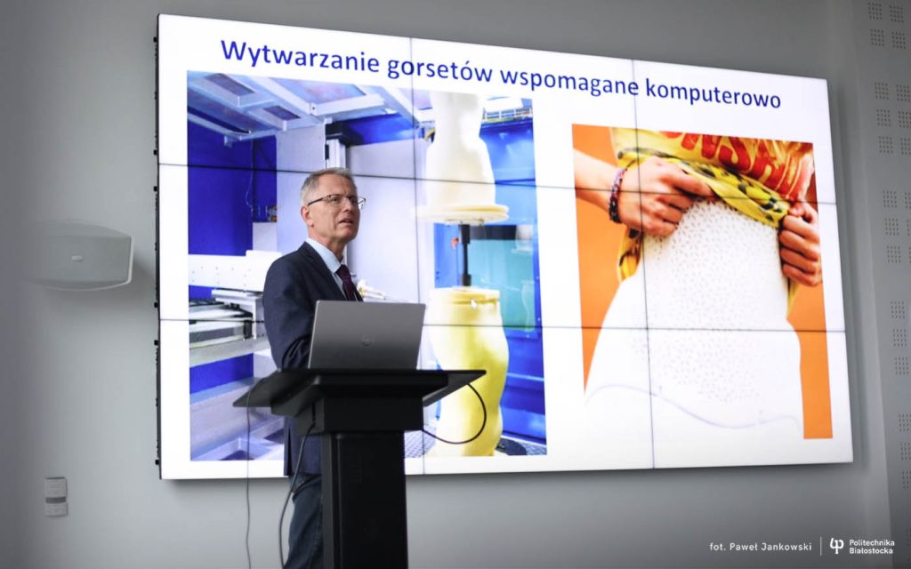
The experiences of such patients drew the attention of Piotr Mrozek, DSc, PhD, Eng., from the Institute of Biomedical Engineering at the Faculty of Mechanical Engineering of Bialystok University of Technology. They inspired a new approach to the traditional method of designing braces.
– We first correct the patient’s trunk while they are positioned in our apparatus to restore proper spinal alignment, and only then do we take measurements, – explains Prof. Mrozek. – The patient is actively involved in the process and provides immediate feedback on the comfort of the correction, while we can observe the effect. Classical methods simply do not allow this.
The corrective apparatus, the key element of the ready-to-implement brace design system, has been granted Class I medical device status.
Patient-Centered Approach
Traditional brace design relies on plaster casts to measure the trunk while the patient is in a relaxed position. Modern methods use 3D optical scanners, but the wet plaster process is time-consuming and uncomfortable for patients.
– During adolescence, changes happen very quickly, – says Prof. Mrozek. – Children aged 10–13 can grow up to 10 cm in a year. Sometimes, two braces may be needed within that period. Any inaccuracies in measurement or initial brace geometry can affect the entire treatment process. For years, we observed how braces were made for children and adolescents with idiopathic scoliosis and saw how burdensome the process was for both patients and orthotic staff.
In collaboration with the Pediatric Orthopedics and Traumatology Clinic of the L. Zamenhof University Children’s Hospital in Bialystok and the “Skoliort” Orthopedic Supply Laboratory in Bialystok, Prof. Mrozek decided to rethink the process. The key question was: “Can the sequence of actions be changed so that trunk correction is planned with the active participation of the patient?”
Active Patient Participation
In classical methods, trunk correction is planned after the fact, based on casts or scans. Patients do not participate in decisions about brace shape, pressure distribution, or tolerance until they try the almost finished brace.
– We wanted to reverse this logic, – explains Prof. Mrozek. – To see if correction could be safely applied first, and only then gather data for the brace design. This transforms the patient from a passive recipient into an active participant in the design process.
This led to a medical experiment concept, with the patient’s active participation as a central element. The study primarily involves patients aged 13–15 with idiopathic scoliosis. The procedure gradually applies trunk correction while monitoring comfort and tolerance limits. Adjustable elements of the apparatus reproduce the pressures on the trunk exactly as the finished brace will.
Only after achieving the optimal trunk alignment is a 3D scan performed. The procedure takes approximately 15–20 minutes, with scanning lasting about two to three minutes. In this system, scanning is a consequence of correction, not the starting point. All actions are performed by a specialist from the orthopedic laboratory.
– Patient feedback is very important to us,- emphasizes Prof. Mrozek. – Despite initial uncertainty, the new procedure does not cause negative reactions. The correction is applied gradually, and the patient can influence its extent. This reduces stress and provides data that were previously unavailable.
Tolerance to correction was found to be highly individual. For the designer, this information is invaluable, allowing better planning of brace geometry digitally. Classical methods lack such a step entirely.
From Correction to Design
In this approach, scanning is not the beginning of the process but its consequence. Data for the brace design are collected after the patient’s correction is applied and accepted.
– The biggest change is the sequence of actions, – explains Prof. Mrozek. – In our system, we first correct the trunk and then perform the scan. Classical methods measure first and apply correction later during the design phase. Here, the patient actively participates throughout the process.
The collected data are input into a computer-aided design system. Reverse engineering is used to create a digital model of the patient’s trunk based on the 3D scan, including precisely planned pressure zones. At this stage, the geometry of the entire brace is designed.
– Using computer methods improves the quality of the design and, consequently, the finished brace, – emphasizes Prof. Mrozek. – We are moving from a process based mainly on orthotist experience and intuition to a system that provides greater control over key parameters.
CNC technology, i.e., numerically controlled machines, allows forming the brace shell exactly according to the digital model. The model shape is reproduced in rigid polyurethane foam, which is milled into a mold used to manufacture the brace shell through thermoplastic methods.
At every stage, a specialist from the cooperating orthopedic laboratory is involved, responsible for design, final production, and patient fitting. The three-way collaboration model—pediatric orthopedic clinic, university, and orthopedic laboratory—ensures continuity between research, design, and clinical practice.
– The clinic handles diagnosis and treatment assessment, we provide innovative tools for design and digital modeling, and the orthopedic lab develops, manufactures, and fits the brace, – explains Prof. Mrozek. – Each element is equally important and directly affects therapeutic outcomes.
Results and Benefits
From the patient’s perspective, the finished brace does not differ significantly in appearance or weight from previous braces. The change is subtle visually but noticeable in daily use.
– The difference lies in design precision and the quality of input data, – notes Prof. Mrozek. – Even after wearing the brace for the first time, we observe a clear reduction in the Cobb angle, the primary indicator of spinal deformity in scoliosis.
Therapy outcomes depend on many factors, including consistent brace use, rehabilitation, and individual patient characteristics. However, better initial fit increases the likelihood of achieving stable results.
Specialists designing and producing braces also benefit. Digital design simplifies the process, shortens preparation time, and allows better control of parameters. Importantly, some design decisions can now be based on objective data rather than solely on specialist experience.
– In traditional methods, much potentially available information is not recorded, – adds Prof. Mrozek. – We collect and use this data during the design phase, improving the overall process quality.
From Research to Implementation
The project is funded by the European Funds for Podlaskie program, with a total budget of PLN 106,807, of which PLN 95,850 covers project implementation. The project leader is Piotr Mrozek, DSc, PhD, Eng., head of the Biomechatronics Department at the Faculty of Mechanical Engineering of Bialystok University of Technology.
– We consider the current system a stage of development, – emphasizes Prof. Mrozek. – We are working on solutions that will further improve brace aesthetics and user comfort in the future.
The solution allows better use of treatment time during the patient’s growth period and reduces the risk of insufficient initial correction. The corrective apparatus has also been submitted for patent protection. The system is ready for clinical use and market introduction.
Compiled by Katarzyna Kozioł


